
Below are listed a variety of protocols designed to yield information about tissue and responses to test compounds and conditions in a range of contexts. Each of these Applications can be performed by EnTox for customers as a Fee for Service. Getting preliminary data is a good way to evaluate whether the BaroFuse is the appropriate instrument to address your research and development needs. Contact EnTox Technical Support to discuss selection of protocols and experimental design.
Drug Toxicity Analysis
Dose Responses
An important application of the BaroFuse is to quantify toxicity of drugs. Dose responses can be characterized by sequentially increasing concentrations of test compound if the affect is less than an hour or so (Fig. 1). Otherwise a time course for each concentration can be assessed individually (Fig 2).
An important application of the BaroFuse is to quantify toxicity of drugs. Dose responses can be characterized by sequentially increasing concentrations of test compound if the affect is less than an hour or so (Fig. 1). Otherwise a time course for each concentration can be assessed individually (Fig 2).
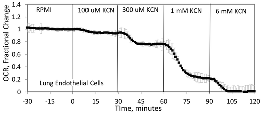
Fig. 1. Cyanide concentration dependency of OCR by Lung Endothelial Cells.
|
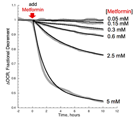
Fig. 2. Metformin concentration dependency of OCR by rat liver slices.
|
Time Courses
In conjunction with the pharmacokinetics of a drug, knowledge of the time course of action on various tissue types. Few commercially available systems can measure slow kinetics of drug response. The BaroFuse can inform regarding the periods of activity subsequent to exposing the tissue to the drug.
In conjunction with the pharmacokinetics of a drug, knowledge of the time course of action on various tissue types. Few commercially available systems can measure slow kinetics of drug response. The BaroFuse can inform regarding the periods of activity subsequent to exposing the tissue to the drug.
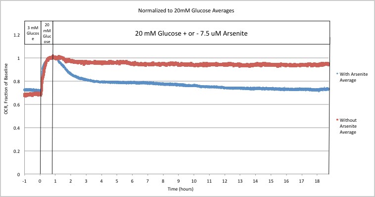
Fig. 1. Acute Drug Effect: Arsenite on Islet OCR.
|
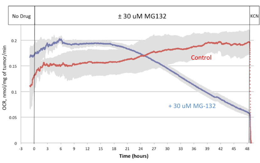
Fig. 2. Delayed Drug Effect: MG-132 on needle biopsy tissue from a mouse tumor made from MDA-MB-231 cells.
|
Metabolic responses
Fuels and drugs that stimulate function and/or metabolism will result in increased OCR. Dose responses can be characterized by sequentially increasing concentrations of test compound if the affect is less than an hour or so (Fig. 1). Otherwise a time course for each concentration can be assessed individually (Fig 2).
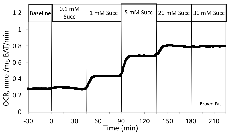
Fig. 1. Succinate concentration dependency of OCR by Brown Adipose Tissue.
|
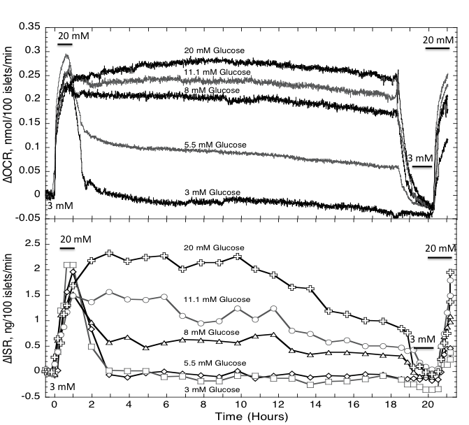
Fig.2. Glucose concentration dependency of OCR by Pancreatic Islets.
|
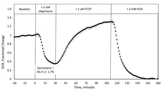
Mitochondrial Stress Test Protocols
Cell Growth Rate / death rate
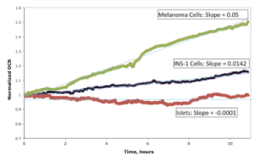
Measuring the rate of change of cell number is commonly done by counting cells, a painstaking and imprecise method that can typically only resolve changes over 48 hours or more. Using the BaroFuse allows for automated measurement of cell growth over short periods of time (Fig. 1), greatly facilitating the process of optimizing growth conditions, and testing drugs or treatments designed to prevent growth or kill the cells. The BaroFuse is the perfect instrument for measuring tissue or cell mass when one does not have access to the specimen such as when designing and optimizing encapsulation devices. In addition, the growth rate can be a robust measure for QA for release of biological products.
If metabolic rate/cell is changing over the time window of assessment then its contribution will have to be distinguished from that made by changing number of cells. Nonetheless, comparisons between cell counting and OCR measurements show that for many situations, changing OCR strongly reflects rate of growth.
If metabolic rate/cell is changing over the time window of assessment then its contribution will have to be distinguished from that made by changing number of cells. Nonetheless, comparisons between cell counting and OCR measurements show that for many situations, changing OCR strongly reflects rate of growth.
|
Fig. 1. Real time determination of cell growth by fast and slow growing cells. Primary islets confirm that OCR is stable when no growth is occurring.
|
Stress tests - Protective treatments
HYPOXIA AND ISCHEMIA / REPERFUSION PROTOCOL
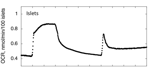
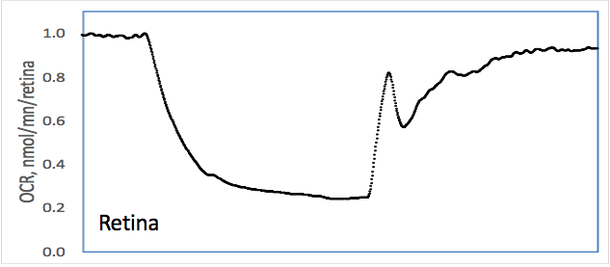
The BaroFuse has the unique ability to measure OCR while changing O2 levels. This feature can allow assessment of tissue sensitivity to periods of low oxygen. Retina, a tissue that is typically functioning at low levels of O2, can completely recover following low O2. In contrast, islets, a micro-organ that relies on metabolic signals to secrete the appropriate amount of insulin, loses almost 50% of pre-hypoxic OCR. The system is therefore suitable to identify drugs and treatments that prevent damage during ischemia, and/or reperfusion.
The BaroFuse has the unique ability to measure OCR while changing O2 levels. This feature can allow assessment of tissue sensitivity to periods of low oxygen. Retina, a tissue that is typically functioning at low levels of O2, can completely recover following low O2. In contrast, islets, a micro-organ that relies on metabolic signals to secrete the appropriate amount of insulin, loses almost 50% of pre-hypoxic OCR. The system is therefore suitable to identify drugs and treatments that prevent damage during ischemia, and/or reperfusion.
|
Fig. 1. Real time OCR in response to a period of hypoxia and return to normoxia. Retina recover, but islets don’t.
|
Effect of dissolved gases
GAS SIGNALLING: H2S, CO, AND NO
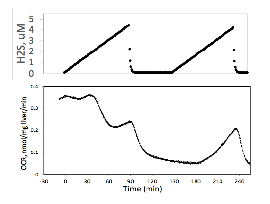
The BaroFuse has the unique ability to measure OCR while changing levels of dissolved trace gases H2S, NO and CO. O2 levels. This feature can allow assessment of tissue response to drugs under regulation from these gas signals. By use of permeation tubes, a known rate of the trace gas accumulates in the BaroFuse and the OCR response is determined as a function of H2S concentration and time of exposure (Fig 1).
The BaroFuse has the unique ability to measure OCR while changing levels of dissolved trace gases H2S, NO and CO. O2 levels. This feature can allow assessment of tissue response to drugs under regulation from these gas signals. By use of permeation tubes, a known rate of the trace gas accumulates in the BaroFuse and the OCR response is determined as a function of H2S concentration and time of exposure (Fig 1).
|
Fig. 1. Effect of H2S on OCR by rat liver. H2S appears to inhibit OCR at low concentration, but then increases at higher concentration when H2S becomes a donor of electrons to the electron transport chain.
|
Direct drug effects
High Resolution analysis of drug effects and time course of action (IC 10, IC 20, IC 50; minutes to 48 hours)
|
Evaluate potential off-target effects: compare IC10 to Cmax
Supplement Pharmacokinetic Characterization (rapid vs. slow responses) |
Rank Magnitude of Effects (2-16 compounds)
|
Selection of Lead Candidates that have least off-target effects
Evaluation of Synthetic Libraries designed to retain efficacy but lose off-target effects |
Rank Tissue Drug Sensitivity (2-14 tissues)
|
Identify tissue most likely to display off-target effects
Maximize efficacy of cancer drugs vs. minimizing damage to normal tissue |
Assess effects of treatment of animal models on drug effects
|
Protein knockouts or protein overexpression
Diet, exercise, gender, age, etc. |
Assess interactions of drug and culture conditions on drug effects
|
Effect of other co-administered drugs and alcohol
Effect of supply fuels (glucose, amino acids, fatty acids) on drug effects |
Biopsied Tissue
|
Individual responses of drugs
|
|
Tumors
Liver Kidney |
Direct metabolic Effects
Evaluate effects of fuels on metabolism
|
Immediate vs. chronic effects
Comparison between sources of energy (glucose, amino acids, fatty acids, ketone bodies) |
Compare Tissue Responses to Fuels (2-14 tissues)
|
Compare OCR vs. Lactate production
|
Assess effects of treatment of animal models on effects of fuels
|
Protein knockouts or protein overexpression
Diet, exercise, gender, age, etc. |
Assess fuels and test compounds on scarce tissue
|
Effect of secretogogs on insulin secretion vs. OCR
Effects on carotid bodies Needle biopsied tissue |
|
Patient studies
Animal models |
Quality Assurance for Release of Tissue
|
Response to fuels, hormones or drugs
Cell Growth Rate Meet criteria for transplantation |
Encapsulated Tissue or Controlled Drug Release devices
|
Stable OCR and/or kinetic profiles of release of drug
|
Stress tests - protective treatments
Stresses: evaluation of OCR before and after the stress
|
Fatty Acids
Cytokines Thapsigargan Oxidative Stress (H2O2) Hypoxia |
Dissolved Gases
Varying Steady State O2 levels
|
Treatments effecting cell growth
On drug toxicity and sensitivity Tissue-specific effects on drug toxicity Effects on VEGF production and other O2-sensitive processes |
Acute Changes in O2 levels
|
Ischemia-Reperfusion Injury
|
Acute Changes in H2S
|
Effects on glycolytic and oxidative metabolism
Protective effect of H2S on stress tests Regulation of insulin secretion |